Mack Lab
Role of Glial Cells in Water and Ion Homeostasis and in Growth Processes in the Adult Central Nervous System
The neuronal environment is largely determined by glial cells regulating the water and ionic homeostasis. We study these cells in various systems from specialized ependymal cells in the human choroid plexus to astroglial cells in fish.
Contact
Andreas Mack, PhD
Working Group Leader
Key Methods of the Lab
Immunocytochemistry, confocal microscopy, electron microscopy, cell and tissue culture
Future directions of the Lab
In a continuously growing nervous system such as the brain and retina of fish, existing cells have to adjust to the changing conditions. We are investigating astroglial cells in brain and retina of teleost fish.
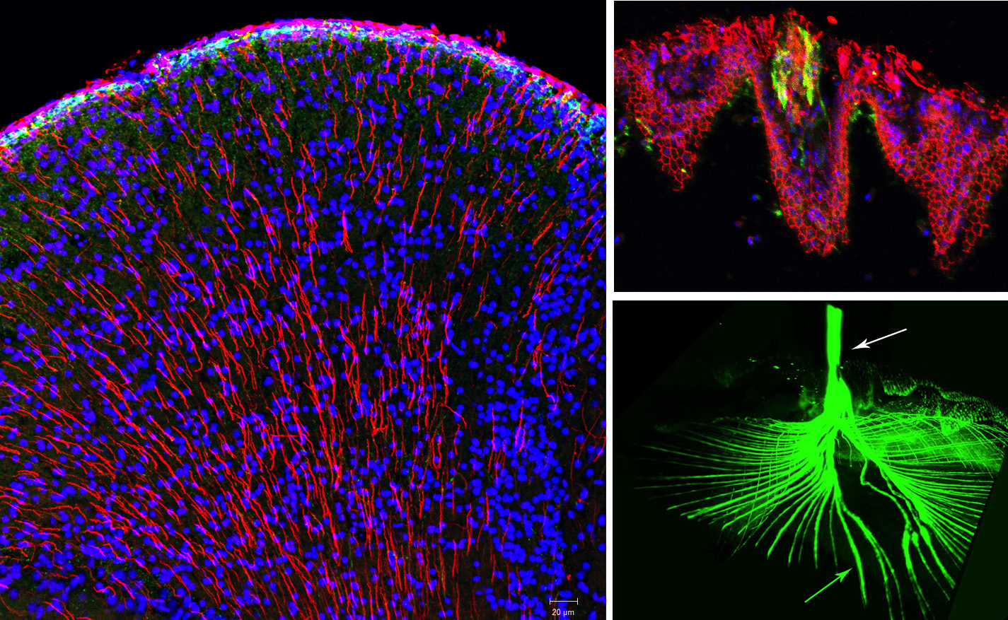
Astroglial cells in the brain of fish have mostly a radial morphology, as shown here in the everted telencephalon of a cichlid fish (left) stained for glial fibrillary acidic protein (red) and maturing neurons labeled with doublecortin (green; see DOI: 10.1002/cne.25126). Thus, radial glial cells are functional glial and dividing progenitor cells at the same time. The cerebrospinal fluid surrounding the ventricular surface is likely produced by cells of the dorsal sac (top right). In addition, we are investigating growth-related processes, structural changes, and neuronal and glial cell additions occurring in the fish retina and optic nerve, where new axons continuously find their way to the brain (bottom right, labelled with doublecortin (in collaboration with Dr. L. deOliveira-Mello, University of Salamanca/ University of Helsinki DOI: 10.3390/biology11020248).
In another line of experiments, we are studying water channels in the ependyma and epithelium of the human and mouse choroid plexus.
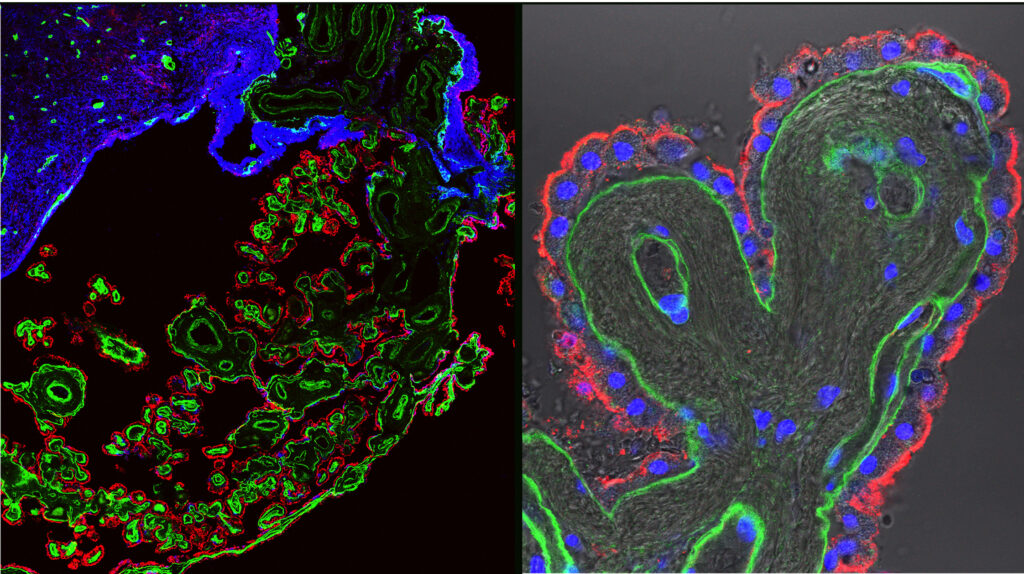
Human choroid plexus stained for aquaporin-1 (red) located apically, and laminin (green) of the basal lamina.
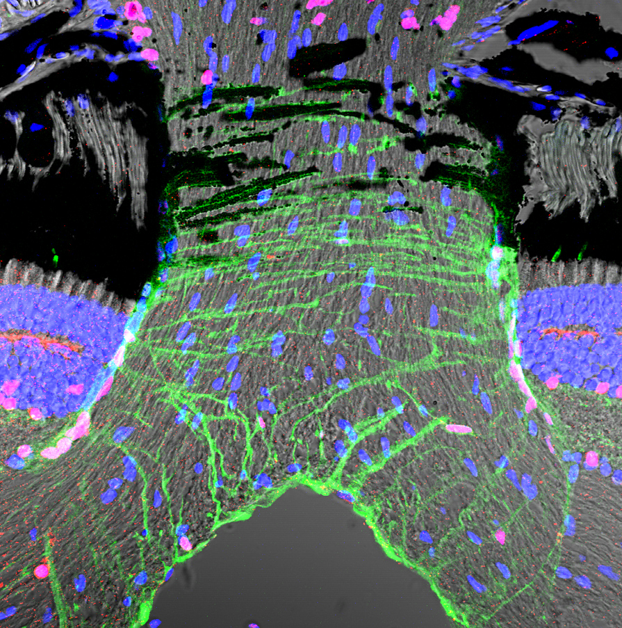
We have recently found that human choroid plexus epithelial cells express the water channel aquaporin-4, in addition to the well documented aquaporin-1 and other channels and transporters. Moreover, we identified a subependymal glial attachment plate with strong aquaporin-4 expression. (see DOI:10.1007/s00018-022-04136-1 and DOI: 10.3390/biom13020212 .
Lab Members
Andreas Mack, PhD
Working Group Leader
Ronja Bihlmaier
Medical PhD Student | IZKF-Promotionskolleg Beneficiary